A Day In the Life of a Biopsy
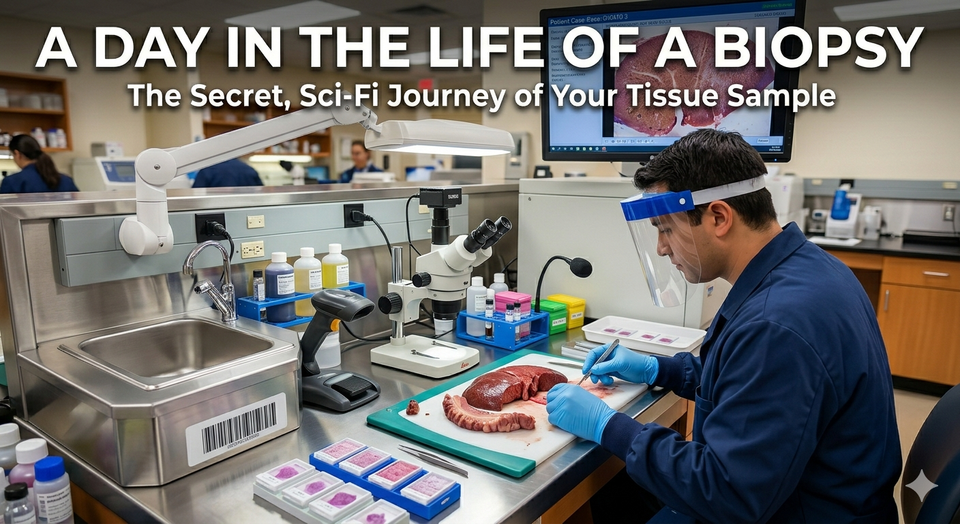
Have you ever wondered what happens after a doctor clips off a tiny piece of tissue from a patient's body and says, "We're going to send this to the lab"? Does it just sit in a jar on a dusty shelf? Does it get popped into a magical machine that instantly blinks a red or green light?
Welcome to the hidden world of the Pathology Department. It is part CSI, part high-tech cooking show, and part elite detective agency. Every single day, hundreds of thousands of tissue samples (often called biopsies) embark on an epic, multi-step adventure. Let’s follow one brave little sample on its journey from a surgical suite to a definitive diagnosis.
Step 1: The Formalin Spa (The Departure)
Our journey begins the moment a surgeon or dermatologist snips a piece of your body off. This might be a mole or a skin tag on the outside of the body; or it might be a polyp or a nodule from the inside! Immediately, the sample is dropped into a plastic cup filled with a pungent liquid called formalin.
But wait! This isn't just a bath; it’s a preservation miracle. When left to its own devices, tissue degrades quickly. Formalin steps in like a biological pause button, "fixing" the cellular structures in place so they look exactly the same weeks, months and even years later.
Step 2: Accessioning (Getting a Passport)
When the sample arrives at the pathology lab, it gets its official identity. In medicine, mixing up samples is the ultimate sin. Therefore, the Accessioning Technician acts as a border control officer.
They check the patient's name, assign a unique barcode, and log it into the computer system. From this moment on, our sample is tracked electronically at every single workstation. No barcode, no entry!
If you are hyper-organized, have an eye for detail, and love data entry with high stakes, this entry-level role is an excellent foot in the door to healthcare.
Step 3: Grossing (The High-Tech Cooking Show)
Don't worry, "grossing" doesn't mean disgusting—it stands for "gross examination," meaning looking at something with the naked eye. This happens at a stainless-steel station equipped with scalpels, rulers, and dictation microphones.
The laboratory professional describes the sample aloud: "Received a 1.2 cm fragment of irregular, tan-pink tissue..." They then skillfully slice it into pieces small enough to fit inside a plastic mesh container called a cassette (about the size of a book of matches).
PAs are the master chefs of the gross room. They have a Master's degree, an incredible grasp of human anatomy, and excellent hands-on surgical skills. They handle everything from tiny moles to entire amputated limbs.
Step 4: The Overnight Cruise (Tissue Processing)
Human tissue is full of water. Unfortunately, water is soft, and you can't cut soft tissue into translucent slices. To fix this, the cassette is loaded into an automated Tissue Processor overnight.
This machine takes our sample on a chemical cruise, bathing it in escalating strengths of alcohol to kick out the water, clearing it with xylene, and finally soaking it in warm, liquid paraffin wax. By morning, the wax has completely penetrated the tissue.
Step 5: Embedding (Making a Micro-Ice Cube)
The next morning, a Histotechnician retrieves the wax-soaked tissue. They place it into a small metal mold and pour fresh, molten wax over it.
They carefully orient the tissue using warm tweezers (if you freeze it upside down, the doctor won't see the right side!). The mold is then placed on a cold plate. The wax hardens rapidly, creating a neat little wax block. Our tissue is now trapped perfectly inside a microscopic "ice cube."
Step 6: Microtomy (The Salami Slicer from Outer Space)
Now comes the pure artistry. The wax block is clamped into a machine called a microtome. Think of a deli meat slicer, but infinitely more precise.
The Histotechnician cranks the wheel, and an ultra-sharp steel blade shaves off a ribbon of wax that is only 3-4 microns thick. To put that in perspective, a single human hair is about 70 microns thick. These ribbons are floated on a warm water bath to smooth out wrinkles, carefully scooped up onto a glass slide, and baked in an oven to melt the tissue securely onto the glass.
Part scientist, part artisan. This career requires incredible fine motor skills, patience, and a love for precision. They turn raw tissue into beautiful, microscopic masterpieces.
Step 7: Staining (The Cellular Glow-Up)
Right now, our tissue slide is practically invisible under a microscope because cells are naturally translucent. It needs some color.
The slide goes through a routine automated staining process called H&E (Hematoxylin and Eosin). Hematoxylin stains the cell nuclei a deep, royal purple, while Eosin stains the cytoplasm and connective tissues a vibrant pink. This pink-and-purple palette is the universal language of pathology.
Step 8: The Grand Finale (The Pathologist's Verdict)
Finally, a glass coverslip is glued over the stained tissue, and the completed slide is placed on the desk of the Pathologist—a specialized medical doctor.
The pathologist places the slide under a high-powered microscope. To the untrained eye, it looks like an abstract pink and purple painting. But to the pathologist, it’s a map. They read the architectural patterns of the cells. They notice if cells are crowded, if the nuclei are abnormally large, or if they are invading places they don't belong.
The pathologist dictates a formal Pathology Report detailing exactly what they see, concluding with a definitive diagnosis (e.g., "Benign mole" or "Adenocarcinoma"). This report is sent back to the patient's primary doctor, who uses the diagnosis to determine the exact treatment plan to best serve the patient.
The "Doctor's Doctor." Pathologists go to medical school and complete residency training. They rarely see patients face-to-face, but they make the final diagnostic decisions that guide all of oncology and surgery.
Why This Journey Matters
Every single slide represents a real human being waiting for answers. Behind the scenes, a highly synchronized team of laboratory professionals works around the clock with absolute precision to ensure that journey is fast, safe, and accurate.
If you love biology, enjoy working with your hands, like solving puzzles, and want to save lives without necessarily standing at a patient's bedside, a career in the laboratory might just be your calling!
Thanks for taking this journey with me! Tune in next week, and may your margins always be clear.
Member discussion