Fixation Series #1 -- Why Fixation Matters: The Chemistry of 10% Neutral Buffered Formalin (And Why Your Tissue Isn't Mush)
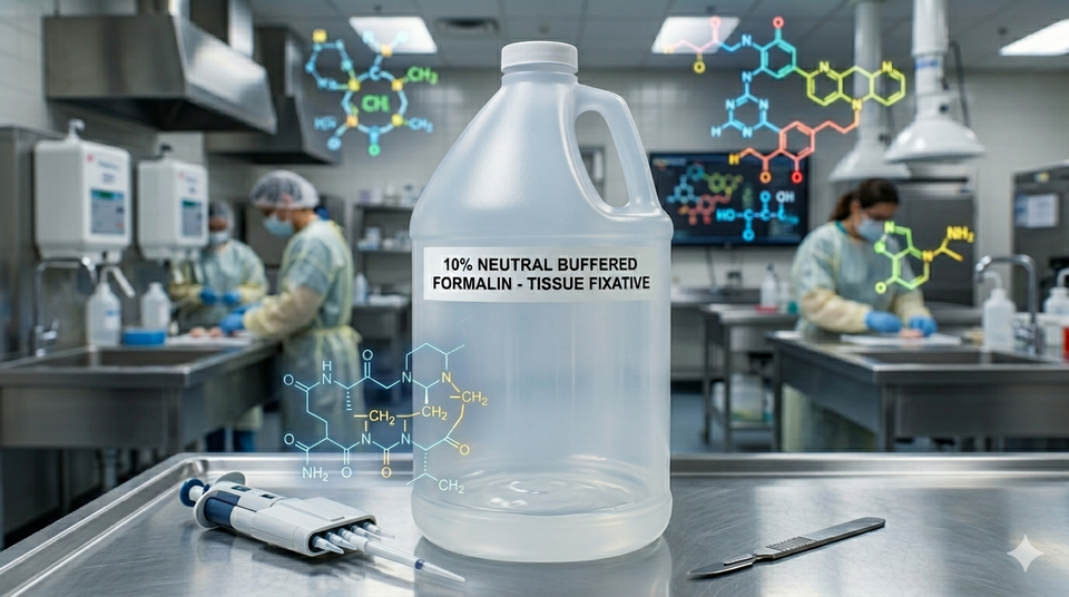
Welcome back, fellow tissue aficionados, to The Formalin Fixation. If you’ve ever stepped into an anatomic pathology lab, you know there is one scent that rules them all. It’s not coffee, it’s not despair—it’s the crisp, wake-up-your-sinuses aroma of 10% Neutral Buffered Formalin (NBF).
Today, we are diving deep into the literal fluid that holds our profession together. We’re talking about the chemistry of fixation. Why do we drown perfectly good tissue biopsies in this pungent cocktail? Because without it, histology would just be a high-stakes game of "guess that puddle."
Autolysis: The Zombie Apocalypse in a Specimen Cup
Before we look at why NBF is the hero of our story, we have to look at the villain: autolysis.
The moment tissue leaves the warm, cozy embrace of the human body, it panics. Deprived of oxygen and nutrients, cells don't just die quietly—they go out in a blaze of self-destructive glory. Intracellular enzymes (specifically from the lysosomes) break free and start digesting the very cell that built them. Left unchecked, your beautifully structured liver biopsy turns into cellular soup within hours.
Fixation is the ultimate pause button. It stops the zombie apocalypse of self-digestion dead in its tracks, preserving the tissue in a state that looks as close to life as possible.
In optimal room-temperature conditions, autolysis begins in highly metabolic tissues (like the kidney or pancreas) within 10 to 15 minutes of blood supply loss. If you don't drop that specimen in fixative quickly, it is literally digesting its own diagnosis.[1]
Enter 10% NBF: The Ultimate Wingman for Proteins
So, what actually is 10% Neutral Buffered Formalin? Let’s break down the recipe.
1. The "Formalin" Part (The Main Event)
First, a bit of mathematical trickery that loves to trip up students on exams: 10% formalin is actually a 4% solution of formaldehyde. Here is how the math shakes out:
- Pure formaldehyde is a gas.
- When dissolved in water to its maximum concentration, it forms a 40% solution called pure formalin.
- We take that pure formalin and dilute it 1:10 with water/buffer.
- Ergo, 10% of 40% = 4% actual formaldehyde gas dissolved in liquid.
2. The Chemistry: Cross-Linking Like a Pro
How does that 4% formaldehyde stop time? It’s all about the methylene bridges.
Formaldehyde (CH₂O) is a highly reactive little molecule. When it penetrates the tissue, it looks for uncharged amino groups on proteins—specifically the amino acid lysine.
The reaction happens in a two-step dance:
- Formaldehyde binds to a nitrogen atom on one protein molecule.
- It then reaches out and binds to a nitrogen atom on a neighboring protein molecule.
This creates a stable, covalent bond known as a methylene bridge (or a cross-link).[2] Imagine a mosh pit where everyone suddenly glues their arms to the people next to them. Nobody can move, nobody can fall over, and nobody can digest themselves. The cellular architecture is locked in place.
Did you know formalin fixation is technically reversible? During immunohistochemistry (IHC), labs use a technique called Heat-Induced Epitope Retrieval (HIER). By boiling the tissue slides in a buffer, they physically break these methylene bridges to uncover hidden proteins. It’s chemical time travel![3]
Why the "Neutral Buffered" Part Isn't Optional
You might be wondering, "Can’t I just mix formaldehyde and tap water and call it a day?" If you want to ruin your pathologist's morning, sure! If you leave formalin unbuffered, atmospheric oxygen will react with the formaldehyde to create formic acid.
As the pH drops, that formic acid reacts with the hemoglobin in your tissue to form a nasty, brown-black crystalline junk known as formalin pigment (or acid formaldehyde hematin). It tends to settle right on top of your cells, mimicking real pathology and masking actual disease.
By adding a sodium phosphate buffer to keep the pH strictly between 7.2 and 7.4, we ensure the formaldehyde does its job without generating a dark, crunchy pigment artifact.[4]
Unbuffered formalin drops the fluid pH down to around 3.5 to 4.5. This acidic environment totally ruins nuclear staining on an H&E slide. If you don't buffer, your tissue will look less like a medical specimen and more like a poorly preserved 3,000-year-old mummy.
By the Numbers: Fun Fixation Facts
To keep your study guides packed, here are a few golden rules of fixation physics:
- The Speed Limit: Formalin penetrates tissue at a rate of roughly 1 mm per hour.[5] If you toss a giant, un-grossed 10 cm tumor into a bucket of formalin, the center will be completely rotten (autolyzed) by the time the fixative finally diffuses to the middle.
- The Golden Ratio: For proper fixation, the volume of your formalin fluid needs to be at least 15 to 20 times the volume of the tissue specimen.[4] Tissue drinks up fixative; don't starve it!
- The Time Warp: Tissue needs to sit in NBF for a bare minimum of 6 to 12 hours for those methylene bridges to properly mature.[5] Take it out too early, and the subsequent alcohol steps in the tissue processor will just dehydrate it into a brittle brick.
The Takeaway
The next time you whiff that unmistakable scent of NBF, give a little nod of respect to the humble methylene bridge. It's keeping autolysis at bay, holding proteins hostage, and ensuring that our slides look sharp enough to make a diagnosis.
Stay fixed, keep your pH neutral, and we’ll see you next time on The Formalin Fixation!
📚 References & Source Material
- Grizzle, W. E. (2009). Special conditions of tissue fixation. Journal of Histotechnology, 32(4), 173-181. (Detailing the rapid onset of cellular autolysis and degradation variables). ↩
- Kiernan, J. A. (2000). Formaldehyde, formalin, glutaraldehyde, and fixation: What are they and what do they do? Microscopy Today, 00(1), 8-12. (The foundational guide to formaldehyde chemistry and lysine cross-linking mechanisms). ↩
- Shi, S. R., Liu, C., & Taylor, C. R. (2007). Standardization of immunohistochemistry for formalin-fixed, paraffin-embedded tissue sections based on the antigen retrieval technique. Progress in Histochemistry and Cytochemistry, 42(3), 127-163. (The definitive review of how HIER undoes methylene bridge cross-links). ↩
- Carson, F. L., & Hladik, C. (2021). Histotechnology: A Self-Instructional Text (5th ed.). ASCP Press. (The gold-standard histology textbook verifying the 7.2–7.4 pH buffering necessity, formalin pigment generation, and the 20:1 fluid-to-tissue ratio). ↩
- Fox, C. H., Johnson, F. B., Whiting, J., & Roller, P. P. (1985). Formaldehyde fixation. Journal of Histochemistry & Cytochemistry, 33(8), 845-853. (Providing classic quantitative data on the distinct rates of formaldehyde penetration vs. cross-linking bound times). ↩
Fixation series #1
Member discussion